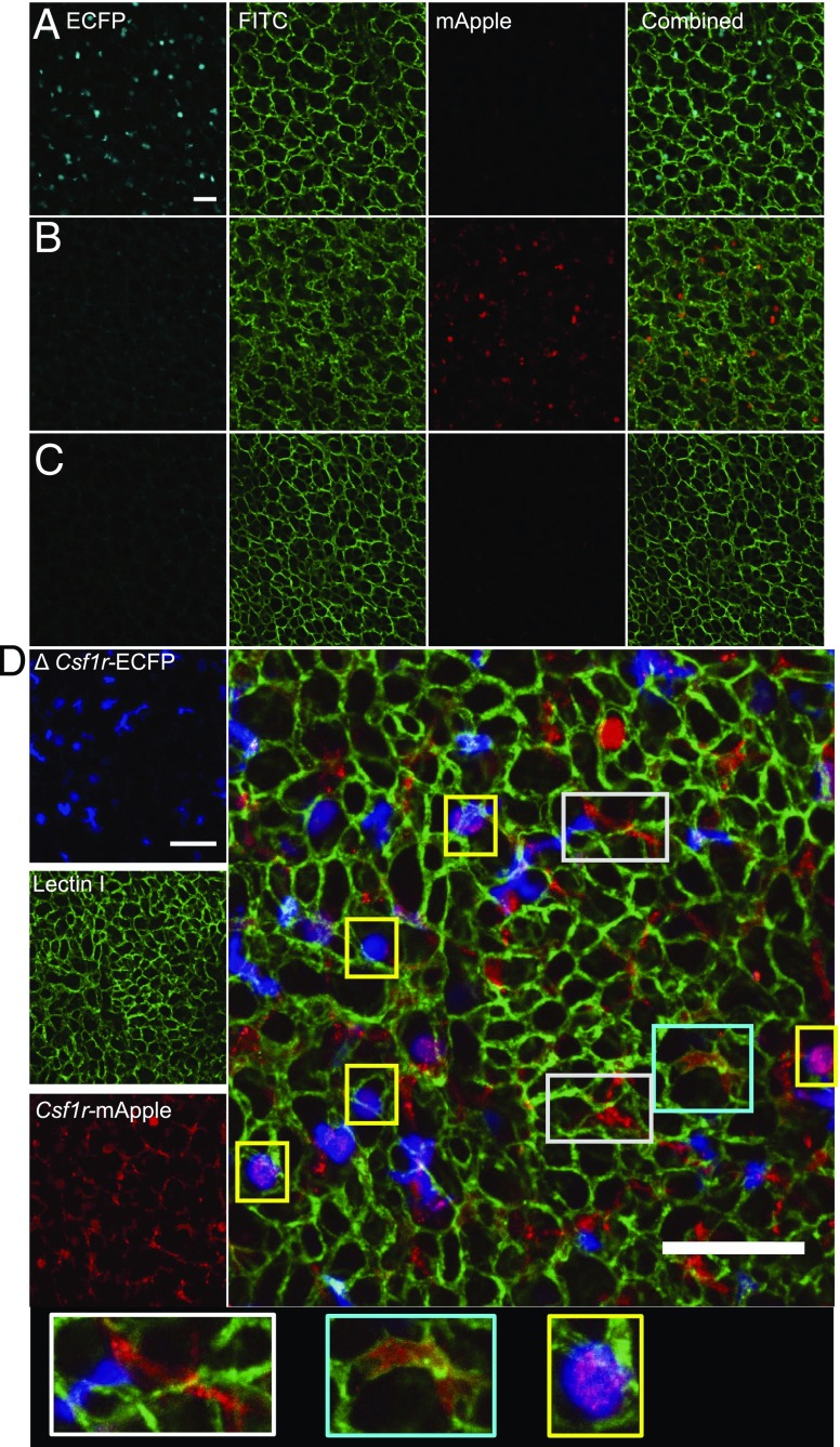
FIGURE 9.
Csf1r-mApple and ΔCsf1r-ECFP transgenes allow imaging of distinct lineages of pulmonary myeloid cells. Confocal image of a transverse section of lung from a ΔCsf1r-ECFP (A), Csf1r-mApple (B), WT (C), and Csf1r-mApple/ΔCsf1r-ECFP (D) mouse imaged ex vivo. FITC-Lectin was injected i.v. to reveal pulmonary vasculature. Scale bars in all panels represent 50 μm.

