
Figure 1.
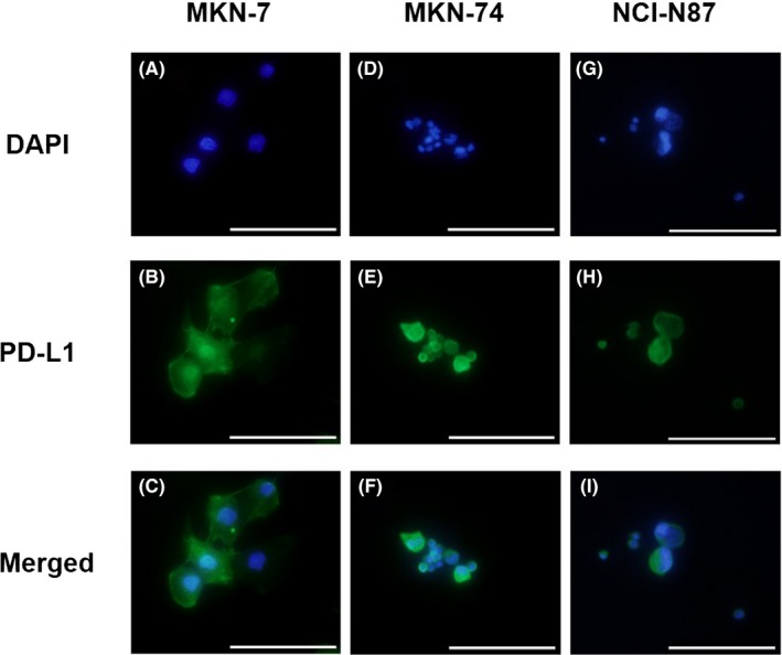
Immunocytochemical staining for programmed death‐ligand 1 (PD‐L1) expression in gastric cancer cell lines using DAPI (A,D,G) and anti‐PD‐L1 mAb–phycoerythrin staining (B,E,H). C,F,I, Merged staining of PD‐L1‐PE and DAPI. PD‐L1 is expressed in tumor cells of MKN‐7, MKN‐74, and NCI‐N87 cell lines. Scale bar = 100 μm (original magnification, ×400)