
Figure 8.
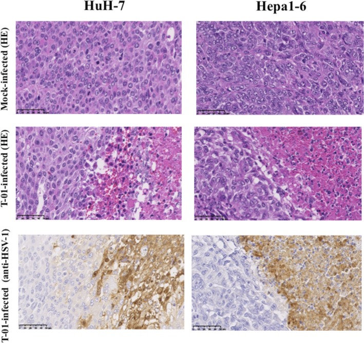
Immunohistochemical analyses of HSV‐1 in mice with subcutaneous tumors. Tumors were induced by subcutaneously injecting HuH‐7 or Hepa1‐6 cells (5 × 10⁶) into the left flank of athymic mice or C57BL/6 mice, respectively. Subcutaneous tumors were treated with mock preparation or T‐01 (2 × 10⁶ pfu) twice (days 0 and 3). Mice were killed 7 d after inoculation, and tissue sections were stained with HE or immunostained with anti‐HSV‐1 antibody. Bar = 50 μm (magnification, ×400)