Fig. 1.

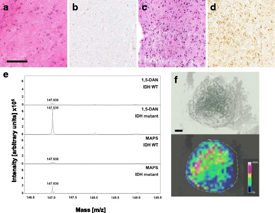
Pre-characterization and 2HG quantification on tissue samples. a H&E staining of IDH wildtype test tissue (CNS-tissue with reactive change), the size bar represents 500 μm. b IDH1R132H staining of IDH wildtype tissue shows no IDH1R132H positive cell. c H&E staining of IDH mutant test tissue (diffuse astrocytoma, WHO grade II). d IDH1R132H staining of test tissue shows IDH1R132H positive cells. e MALDI-TOF spectra showing m/z values from 146.5 to 149.5 of IDH wildtype and IDH mutant tissues with the matrices 1,5-DAN and MAPS. Peaks at m/z 147 correspond to 2HG. f Light microscopic image and corresponding MALDI-TOF image of a representative 0.05 μl 1,5-DAN spot on IDH mutant tissue. The size bar represents 150 μm, the intensity bar shows the color coding for MALDI-TOF intensities within the scanned region
