Fig. 3.

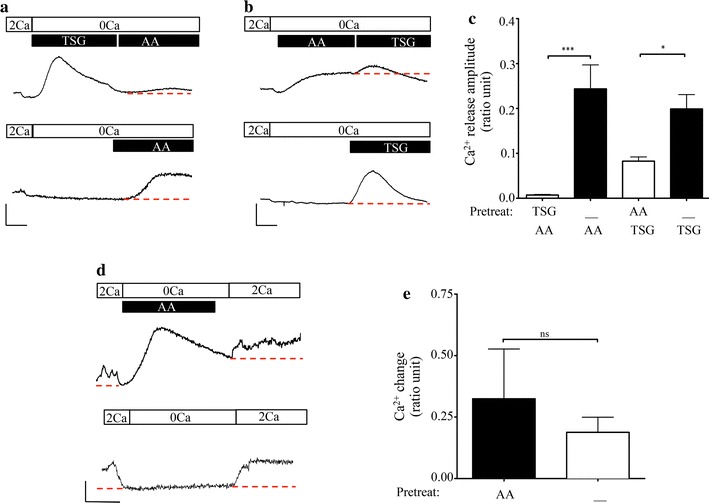
Treatment with AA depleted [Ca2+]ER, but was not sufficient to evoke SOCE. a Upper panel: representative trace showing pre-treatment of fura-loaded BON cells with 1 μM TSG in nominal Ca2+-containing media abolished AA-mediated release. Lower panel: representative time-matched control trace where TSG pre-treatment was omitted. Scale bars indicate 100 s (horizontal) and 0.1 ratio units (vertical). b Upper panel: representative trace showing pre-treatment with 6 μM AA in nominal Ca2+-containing media significantly diminished the TSG-mediated release. Lower panel: representative time-matched control trace where pre-treatment with AA was omitted. Scale bars indicate 100 s and 0.1 ratio units. c Bar chart showing average data for treatments indicated in panels a and b. d Upper panel: representative trace showing the effect of pre-treatment with 6 μM AA in nominal Ca2+-containing media on Ca2+ entry. Lower panel: representative time-matched control trace where pre-treatment with AA was omitted. Scale bars: x-axis = 100 s, y-axis = 0.1 ratio units. e Bar chart showing average change in cytosolic Ca2+ levels in response to change in extracellular Ca2+ concentration. In figures a, b and d magnitude of Ca2+ responses were measured by determining change in amplitude with respect to the red dashed line. Significance of p < 0.05 and p < 0.001 is indicated by * and ***, respectively
