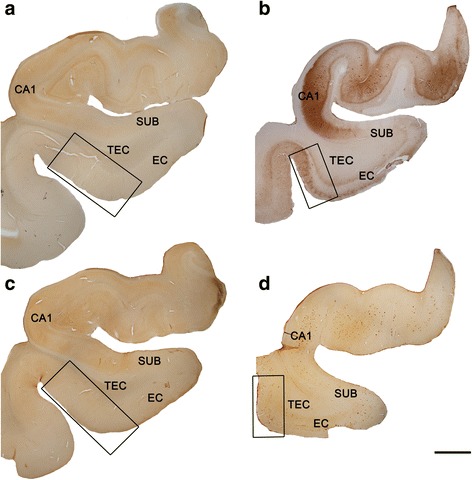
Fig. 2.

Coronal sections of human hippocampal formation. Low-power photographs of a control subject (a, c) and an AD patient (b, d), in sections immunostained for anti-PHF-Tau-AT8 (a, b) and anti-Aβ (c, d). TEC is indicated by the box. Immunostaining for anti-PHF-Tau-AT8 (b) and anti-Aβ (d) can be observed in the AD patient. These neuropathological marks are absent in the control subject (a, c). Scale bar (in d): 3 mm
