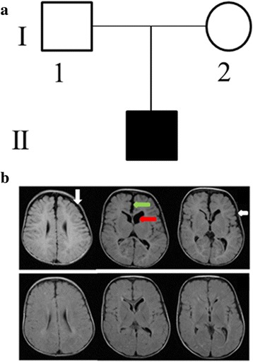
Fig. 1.

Clinical and molecular analysis of an Emirati family with a child affected by CH. a Pedigree showing the affected child with hydrocephalus for a non-consanguineous Emirati family. b MRI (T1 weighted images), upper panels at age 3 months showing widened CSF spaces in the frontoparietal regions (white arrows), widened interhemispheric fissure (green arrow) and mild dilatation of ventricles (red arrow). The lower panels at age of 17 months showing improvement in the above mentioned findings
