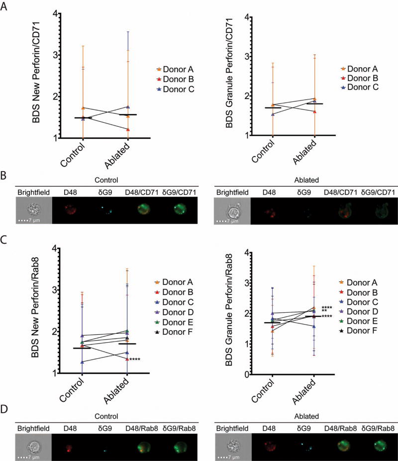
Fig 5. Ablation of recycling endosomes increases perforin localization with rab8.
CD8+ T cells were isolated by negative selection and ablated of their recycling endosome function after stimulation with anti-CD3 and anti-CD28/CD49d for 3 hours. 3–6 separate donors (Donors A–F) were used, with 60–1300 cells analyzed per donor. Median BDS (similarity) scores of new perforin and granule perforin with CD71 (A–B) and rab8 (C–D) are shown, including representative images (B and D). Horizontal lines represent the mean of all donors combined, and error bars show the standard deviation of each donor. **p<0.01, ****p<0.0001, 2 way ANOVA with Holm-Sidak multiple comparisons test between control and ablated cells.

