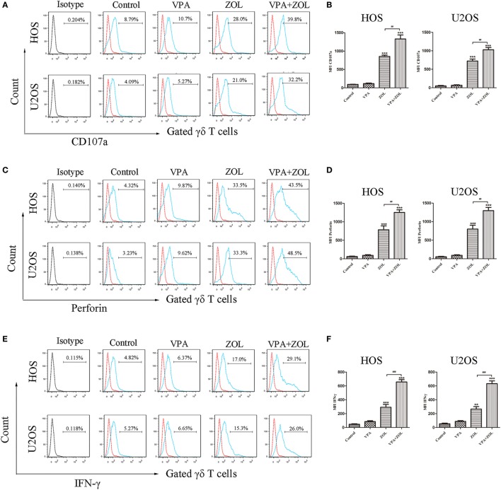
Figure 2.
Valproic acid (VPA) and zoledronate (ZOL) upregulated the level of γδ T cells cytotoxicity-related indicators against osteosarcoma cells. HOS and U2OS cells were treated for 24 h with control, 1 mM VPA, 2 µM ZOL, and VPA + ZOL, respectively, before 2 h co-cultured with γδ T cells at an E:T ratio of 5:1. (A,B) CD107a expression levels on γδ T cells were measured by flow cytometry. MFI of CD107a+ γδ T cells was presented in histograms. (C,D) Perforin expression levels of γδ T cells were measured by flow cytometry. MFI of perforin+ γδ T cells was presented in histograms. (E,F) IFN-γ expression levels of γδ T cells were measured by flow cytometry. MFI of IFN-γ+ γδ T cells was presented in histograms. Red histogram line was isotype control and panels were overlapped. All the values were shown as mean ± SD from three independent experiments; *p < 0.05, **p < 0.01, ***p < 0.001 versus control, #p < 0.05.

