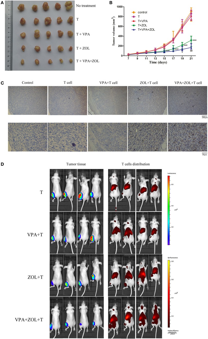
Figure 6.
Valproic acid (VPA) and zoledronate (ZOL) enhanced γδ T cells cytotoxicity against osteosarcoma in xenograft models. HOS cells transfected with luciferase (HOS-Luc) were inoculated subcutaneously into the right flank of BALB/c-nu mice. After 7 days, mice started to receive various treatments and injection of γδ T cells. (A) Tumor excised from mice on 21th day. (B) Tumor volumes were measured every 2 days, starting on the seventh day. (C) Intratumoral γδ T cells were detected by immunohistochemical assays (shown in brown and indicated with arrowheads). (D) Orthotopic models were established using HOS-Luc cells and BALB/c-nu mice. After 7 days, mice started to receive various treatments and injection of γδ T cells. Mice were imaged with in vivo imaging system 24 h postinjection of γδ T cells. Tumor growth was evaluated by visualizing bioluminescence and T cells migration was shown by DiR fluorescence. All the values were shown as mean ± SD; ***p < 0.001 versus control, #p < 0.05, ###p < 0.001 versus treatment with ZOL and γδ T cells injection.

