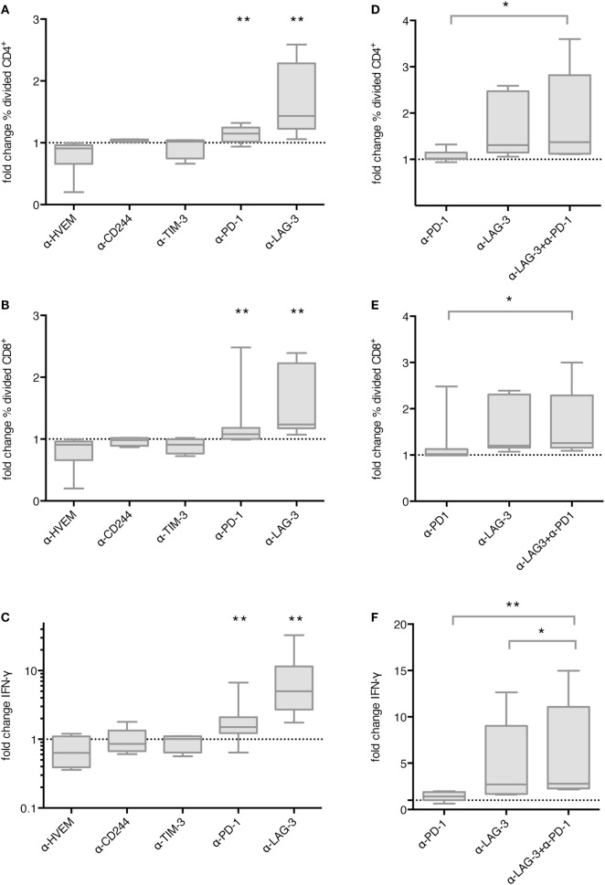
Figure 3.
Effect of immune checkpoint blockade on proliferation and IFN-γ secretion of T cells after stimulation with TLR-3-DCs. CD3+ T cells of 4–14 healthy donor (HDs) were cocultured with autologous CMV, EBV, influenza, tetanus (CEFT)-pulsed TLR-3-DCs in the presence or absence of immune checkpoint blocking antibodies, either for individual antibodies (A–C) or in different combinations of α-PD-1 and α-LAG-3 antibodies (D–F). Proliferation of CD4+ (A,D) and CD8+ T cells (B,E) was analyzed by carboxyfluorescein N-succinimidyl ester (CFSE) assay, and the ratio between the percentages of divided cells with and without blocking antibody was calculated. IFN-γ secretion of CD3+ T cells (C,F) was determined by cytometric bead array (CBA) assay, and the ratio between concentration with and without blocking antibody was calculated. All data are presented as box-and-whisker plots, and statistical significance was calculated against a fold change of 1.0. *p < 0.05; **p < 0.01.

