
Figure 5.
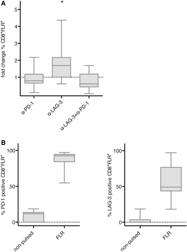
Effect of programmed cell death protein 1 (PD-1) and lymphocyte activation gene 3 (LAG-3) blockade on proliferation of EBV antigen-specific T cells after stimulation with TLR-3-DCs. non-adherent cell (NACs) of 9 healthy donor (HDs) were cocultured with autologous Epstein–Barr nuclear Ag 3 A peptide FLRGRAYGL (FLR)-pulsed TLR-3-DCs in the presence or absence of α-PD-1 and α-LAG-3 antibody. (A) The percentage of FLR tetramer positive cells within the CD8+ T cell population was determined by flow cytometry. Data for fold change to the condition without blocking antibody are presented as box-and-whisker plots, and statistical significance was calculated against a fold change of 1.0. *p < 0.05. (B) PD-1 and LAG-3 expression was determined for FLR tetramer positive CD8+ T cells after stimulation with non-pulsed or FLR-pulsed TLR-3-DCs.