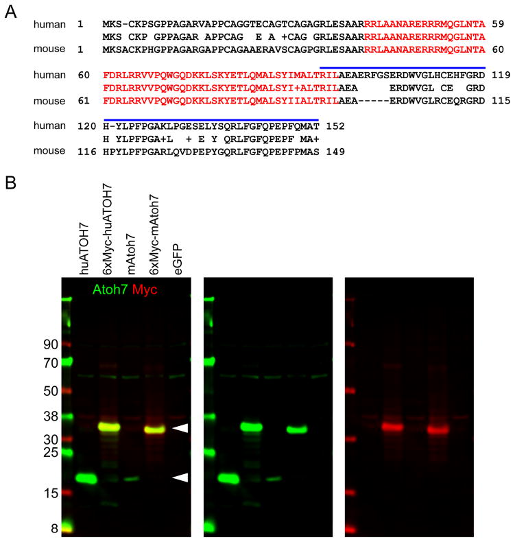
Fig. 1. Detection of human and mouse Atoh7.
[A] Alignment of human and mouse Atoh7 polypeptides. The bHLH domain (red, 56 amino acids) and human recombinant peptide immunogen (blue bar, 59 amino acids) are indicated. The human and mouse sequences are 82% identical overall, 98% identical in the bHLH domain, and 70% identical in the C-terminal segment. [B] Western analysis of HEK293T cells transfected with human (huATOH7) or mouse (mAtoh7) expression plasmids ± six myc epitope tags. Arrowheads indicate the positions of Atoh7 (17 kDa) and 6xMyc-Atoh7 (predicted size, 27 kDa; actual size, ~32 kDa) proteins. The pCS2-mAtoh7 transfection was notably less efficient than other samples. The Atoh7 antibody cross-reacts weakly with a 60 kDa human protein in HEK293T.

