
Figure 1. Examples of corticocortical and thalamocortical functional connectivity changes in anesthesia and disorders of consciousness.
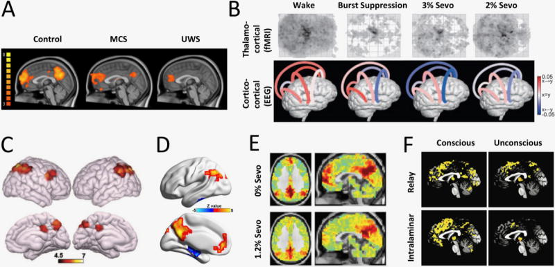
(A) Comparison of functional magnetic resonance imaging (fMRI) connectivity of the default mode network (DMN) in healthy participants, patients in minimally conscious state (MCS), and those with unresponsive wakefulness syndrome (UWS). Partial preservation of the DMN is observed, especially in MCS patients (from Di Perri et al, 2014). (B) Effect of sevoflurane anesthesia on functional connectivity. Top: widespread reduction in fMRI thalamocortical connectivity, especially with frontal cortex at all concentrations of sevoflurane. Bottom: changes in directed connectivity as measured by EEG symbolic transfer entropy. Decreases from frontal to parietal, temporal and occipital cortex and from temporal to parietal cortex are evident. Color encodes the direction of information flow (red: rostrocaudal, blue: caudorostral) (from Ranft et al, 2015). (C) Increased frontoparietal network connectivity in MCS compared to UWS patients. Consistent differences are also found in DMN, salience, and sensory-motor networks (not shown) (from Demertzi et al, 2015). (D) Regions whose fMRI functional connectivity correlate with the Glasgow Coma Scale of conscious, MCS, and UWS patients with acquired brain injury. Positive correlation with the level of consciousness is found in the default mode network (from Wu et al, 2015). (E) Sevoflurane anesthesia (1.2%) functionally ‘disconnects’ medial prefrontal cortex from the DMN (fMRI functional connectivity with posterior cingulate seed). Thalamocortical connectivity with the DMN is also reduced (not shown) (from Palanca et al, 2015). (F) Differential modulation of thalamocortical functional connectivity of the specific relay and the “nonspecific” intralaminar nuclei (fMRI data). Deep sedation with propofol (unconscious) exerts widespread reduction of intralaminar thalamocortical connectivity (from Liu et al, 2013).