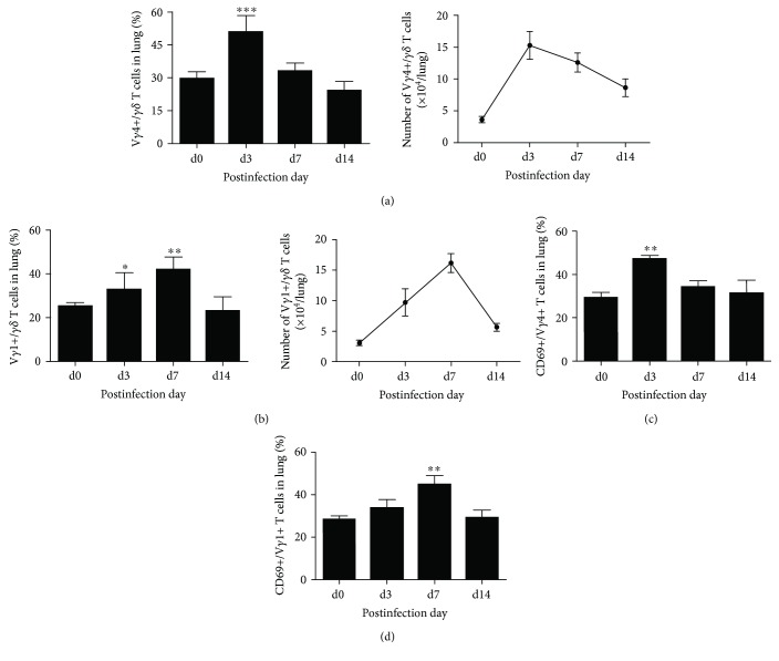
Figure 3.
Vγ1+ T and Vγ4+ T cell proliferated and activated during Cm respiratory infection. Mononuclear cells in lung tissues at different time points postinfection were extracted. Staining with anti-mouse CD3, TCRγδ, TCRVγ1, and TCRVγ4 antibody to analyze the percentage and absolute number of TCRVγ1+ TCRγδ+ T cells (a) and TCRVγ4+TCRγδ+T cells (b) by flow cytometry. The activation extent of Vγ1+ T (c) and Vγ4+ T (d) cell was measured by the expression of CD69, staining with anti-mouse CD69 antibody. The results are presented as mean ± SD. ∗p < 0.05, ∗∗p < 0.01, and ∗∗∗p < 0.001.

