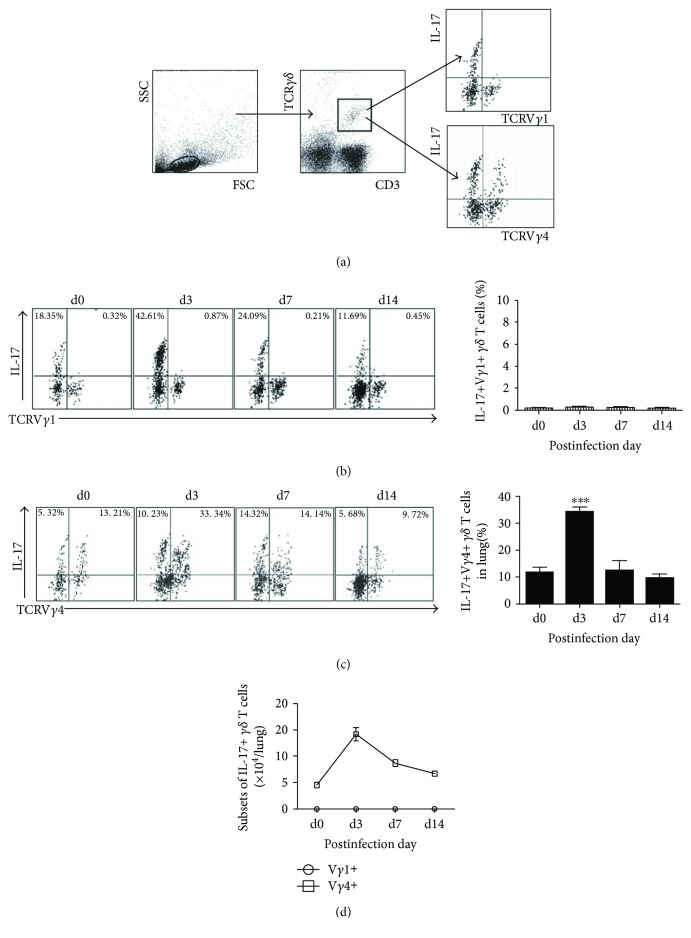
Figure 5.
Vγ4 cells at day 3 p.i. are the major sources of IL-17 during Cm lung infection. IL-17+ Vγ4+/Vγ1+ T cells were gated (a), stained with anti-mouse CD3, TCRγδ, TCRVγ4, and IFNγ/IL-17 antibody to analyze percentage of Il-17+ Vγ1+ T cells (b) and IL-17+ Vγ4+ T cells (c) in lung tissues after Cm infection. Comparison between IL-17+ Vγ1+ cell and IL-17+ Vγ4+ cell with its absolute number (d). The results are presented as mean ± SD. ∗∗∗p < 0.001.

