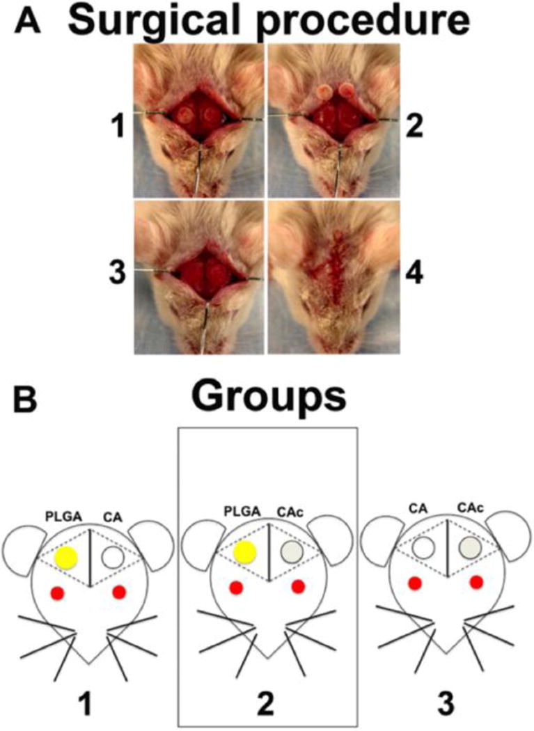
Figure 1. Surgical procedure and schematic of the study groups.

(A) Steps in the surgical implantation: (1) Creation of two circular 3.5mm defects on the two sides of mouse calvaria, (2) removal of calvarial bone, (3) Implantation of the scaffolds into the defects, (4) Closure of the implants by suturing the skin. (B) Groups 1: left side defect was filled with PLGA and the right side defect with CA, group 2: left side defect was filled with PLGA and the right side was filled with CAc, group 3: left side was filled with CA and the right side was filled with CAc. In study 1, the materials alone were used. In study 2, 1×10⁶ Col3.6-Cyan BMSCs from donor mice were seeded on to each scaffold before implantation into host mice with Col3.6-Tpz BMSCs.
