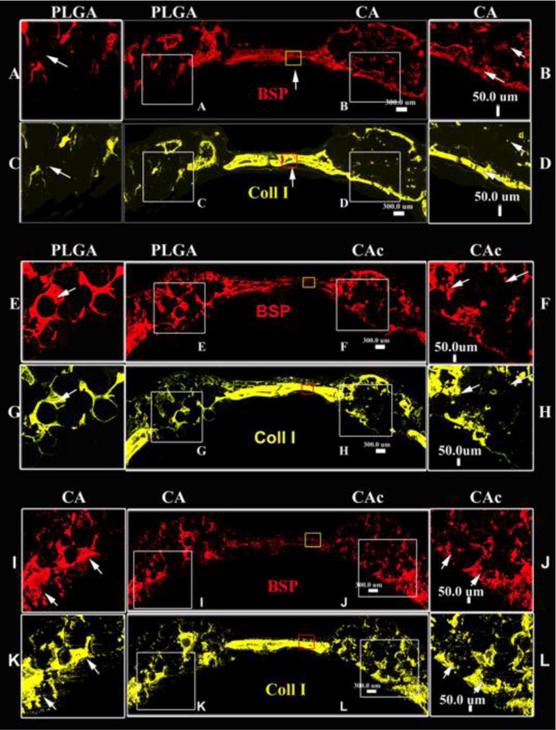
Figure 4. Deposition of ECM proteins with implantation of materials with donor cells.

Fluorescent histological cross sectional images of calvaria implanted with material and cells at 8 weeks, A-D-Group 1, PLGA vs CA; E-H-Group 2, PLGA vs CAc; I-L-Group 3, CA vs CAc; Bone Sialoprotein (BSP) (red), Collagen 1 (yellow) –Bone ECM proteins.
