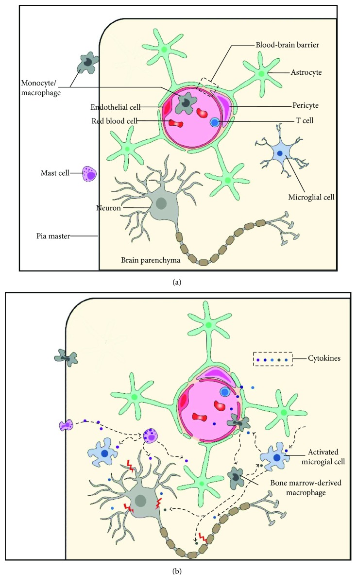
Figure 1.
Schematic diagram of immune cells in POCD. (a) Under a normal condition, neurons are normally functioning. Microglia are ramified and in a resting state. The BBB is intact. Monocytes, mast cells, and T cells are restricted outside the brain parenchyma. (b) After surgery, many cytokines are released from the injured sites and damage the BBB. Microglia are triggered by these cytokines and turned into an activated, amoeboid shape. Microglia-secreted cytokines can damage neurons and also recruit BMDMs and other inflammatory cells from the blood. BMDMs and MCs infiltrate into the brain parenchyma and release more cytokines, which can directly damage neurons and also activate microglia. Cytokines secreted by T cells also participate in neuroinflammation in POCD. The immune cells and cytokines compose an inflammation network that aggravates neural damage, leading to POCD. POCD: postoperative cognitive dysfunction; BBB: blood-brain barrier; BMDM: bone marrow-derived macrophage; MC: mast cell.

