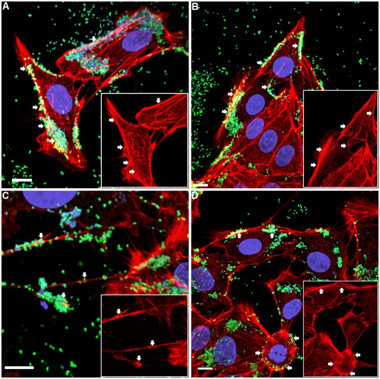
Figure 1.
Confocal micrograph overlays of adherence patterns of M. hyopneumoniae cells to PK-15 monolayers. M. hyopneumoniae cells were labeled with F2P94−J antisera conjugated to CF™ 488 (green), nucleic acids were stained with DAPI (blue), and F-actin was stained using phalloidin (red). M. hyopneumoniae cells can be seen adhering to the edges of the monolayer and to cellular projections that appear to be filopodia (white arrows). Inserts in each panel depict the phalloidin channel of each image (more specifically, the areas highlighted in the overlays), highlighting areas that stain intensely for F-actin (white arrows). (A–D) represents images taken from individual replicate experiments. Scale bars are 10 μm.

