Figure 4.
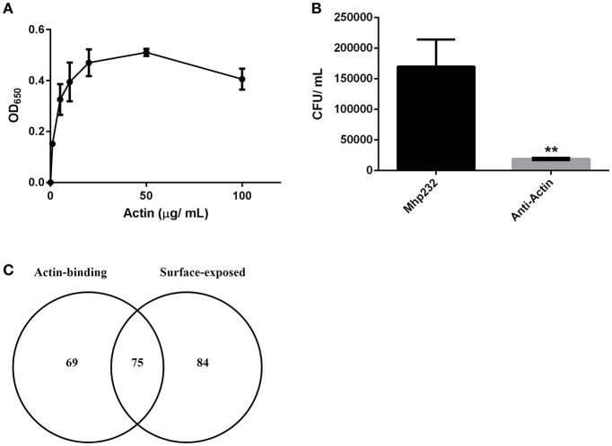
M. hyopneumoniae binds actin. (A) Microtiter plate binding assay of immobilized M. hyopneumoniae cells binding to actin in a dose-dependent and saturable manner. Each data point represents the average of individual triplicate experiments with the standard error of the mean shown. (B) Treatment of PK-15 cell monolayers with mAbs that target actin inhibited adherence of M. hyopneumoniae by ~90%. The data is presented as the average of triplicate experiments and the standard error of the mean. An unpaired t-test was performed with a p < 0.01, indicated as **. (C) Venn diagram showing the overlap between putative actin-binding proteins purified using actin affinity chromatography, and M. hyopneumoniae surface proteins.

