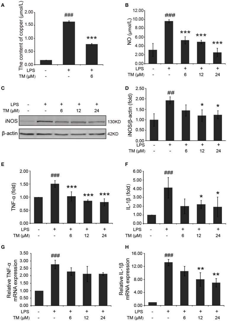
Figure 3.
TM-pretreatment reduces the levels of copper and inhibits the production of NO and the expression of pro-inflammatory cytokines in LPS-induced BV-2 cells. BV-2 cells were pretreated with 6, 12, 24 μM of TM followed by treatment with 1 μg/ml LPS for 18 h. (A) The levels of copper were detected by using the ICP-MS. (B) NO production was detected by Griess agent. (C,D) Immunoblot images (C) and quantifications (D) show that TM-pretreatment suppressed iNOS expression in LPS-induced BV2 cells. (E,F) ELISA assay data show that TM-pretreatment decreased the release of TNF-α and IL-1β in LPS-induced BV2 cells. The absolute values ranges of TNF-α and IL-1β were 1000–2000 pg/ml and 102–993 pg/ml. (G,H) Quantitative real-time PCR (qPCR) data show that TM-pretreatment blocked TNF-α and IL-1β mRNA expression in LPS-induced BV2 cells. Data are represented as means ± SD. of at least three independent experiments (N ≥ 3). ***p < 0.001 **p < 0.01, *p < 0.05 compared with the LPS; ###p < 0.001, ##p < 0.01 compared with the control. The p-values were calculated by One-way ANOVA followed by Bonferroni's post-hoc test.

