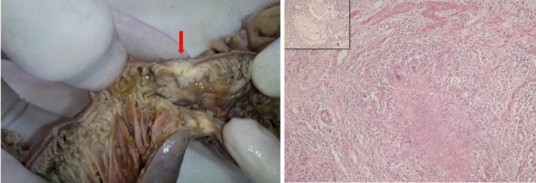
Figure 1.
Left: Segment of small bowel from first laparotomy showing a stricture with luminal narrowing (arrow). Right: Epithelioid cell granuloma with central caseous necrosis and Langhans giant cell seen adjacent to muscularis mucosae (HE-100 x). Inset shows granuloma in mesenteric lymph node dissected from the specimen.

