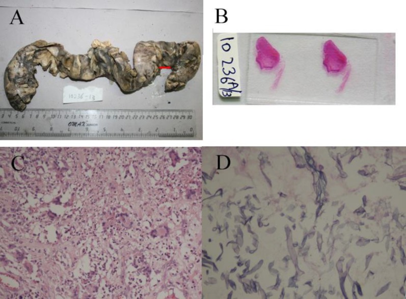
Figure 2.
Segment of small bowel from the second laparotomy showing exudate at several places. A perforation (arrow) is also seen near one of the resected ends. B: Marked wall thinning suggestive of impending perforation as seen on naked eye examination of slide. C: Florid giant cell reaction showing several intra-cellular fungi (HE – 200x). D: High power showing typical fungal morphology of aseptate hyphae, section from a thinned out area (PAS – 400x).

