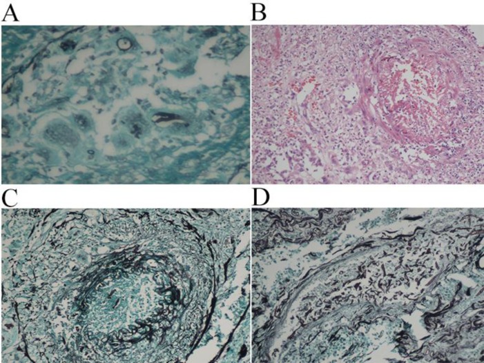
Figure 3.
A: Grocott Gomori methanamine silver stain showing broad fungal hyphae engulfed by a giant cell (GMS – 400x). B: Blood vessel showing vasculitis and lumen obliteration by thrombosis (HE – 200x). C: Grocott Gomori methenamine silver stain showing intra-vascular hyphae (GMS – 200x). D: Grocott Gomori methenamine silver stain highlighting the presence of numerous fungal hyphae inside the lumen of a blood vessel (GMS – 200x).

