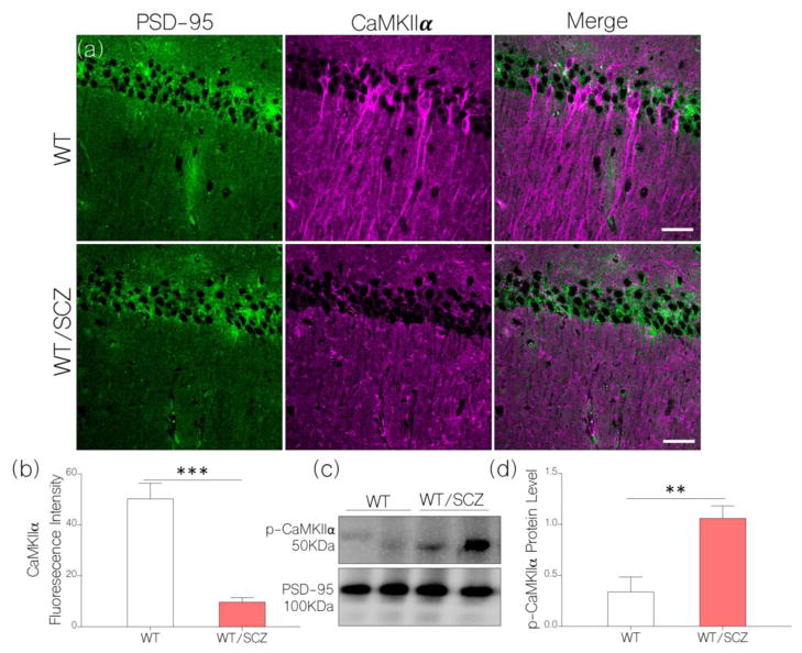
Figure 5.
a, Representative confocal images (scale bar=20μm) show a significant decrease in hippocampal CaMKIIα after NMDAR hypofunction was induced (WT/SCZ) in mice (p<0.001): versus the control (WT).
b, Bar chart comparing the expression of hippocampal CaMKIIα for control (WT) and behaviorally deficient NMDAR hypofunction mice (WT/SCZ).
c, Quantitative immunoblot and d, bar chart shows increased phosphorylation of CaMKIIα in hippocampal lysate of WT/SCZ mice when compared with the control (p<0.01).

