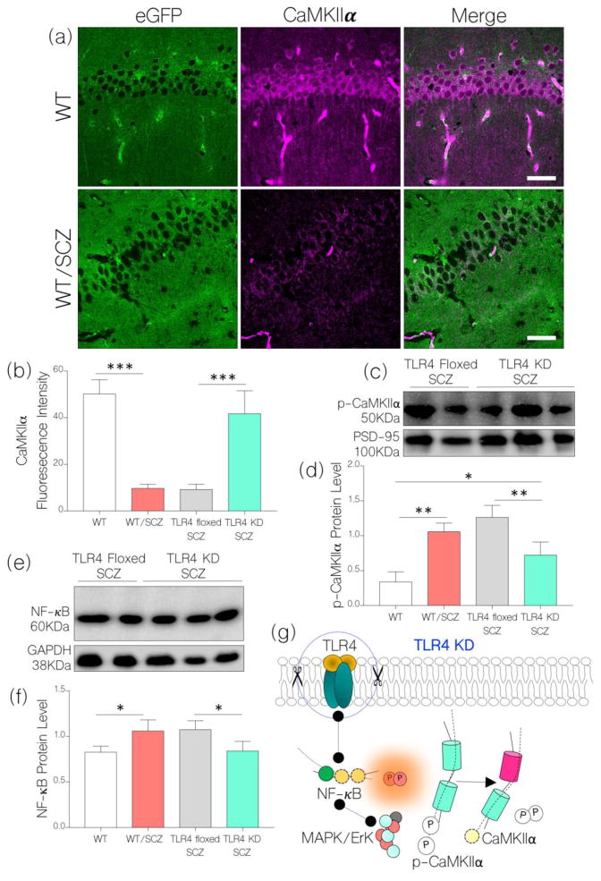
Figure 8.
a, Representative confocal images show sustained expression of CaMKIIα in the hippocampus of TLR4 KD SCZ mice after an induced NMDAR hypofunction. When compared with the WT (control), no significant change in CaMKIIα expression was recorded. Like WT/SCZ, neural CaMKIIα expression reduced significantly in TLR4 floxed SCZ mice when compared with the control (p<0.001).
b, Bar chart illustrating comparative (normalized) expression of CaMKIIα in the hippocampus of WT, WT/SCZ, TLR4 floxed SCZ, and TLR4 KD SCZ mice (One-Way ANOVA).
c, Western blot and d, bar chart showing a change in phosphorylated CaMKIIα (p-CaMKIIα) in hippocampal lysate of TLR4 KD SCZ and TLR4 floxed SCZ mice.
e, Immunoblot and f, bar chart shows a reduced NF-κB expression in hippocampal lysate of TLR4 KD SCZ when compared with TLR4 floxed SCZ mice (p<0.05). No significant change in NF-κB was recorded for the TLR4 KD SCZ group when compared with the control (WT).
g, Schematic illustration of a possible mechanism through which TLR4 knockdown might have attenuated hippocampal CaMKIIα loss and restored NMDAR function in schizophrenia.

