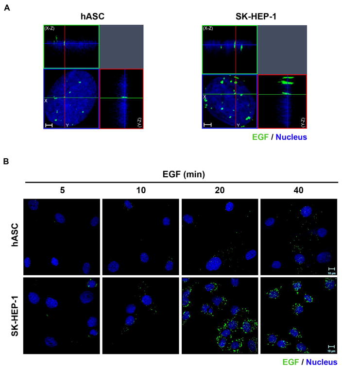
Fig. 2.
EGF-488 clusters translocate to the nucleus in hASC and SK-HEP-1 cells. A- Three-dimensional reconstruction of optical sections of the hASC and SK-HEP-1 were performed, after cells were stimulated for 40 min with EGF conjugated with Alexa Fluor® 488 (in green). X–Z sections are shown at the top, and Y–Z sections are shown on the right of each image. Images are representative of 41 (hASC) and 37 (SK-HEP-1) cells. Nuclei are in blue. Scale bar: 2 μm. B- Cells were stimulated with EGF conjugated with Alexa Fluor® 488 for 5, 10, 20 and 40 min (in green), had their nuclei labeled with Hoechst (in blue) and were analyzed by super-resolution microscopy. Images are representative of 8 to 10 fields. Scale bar: 10 μm. (For interpretation of the references to color in this figure legend, the reader is referred to the web version of this article.)

