Fig. 5.
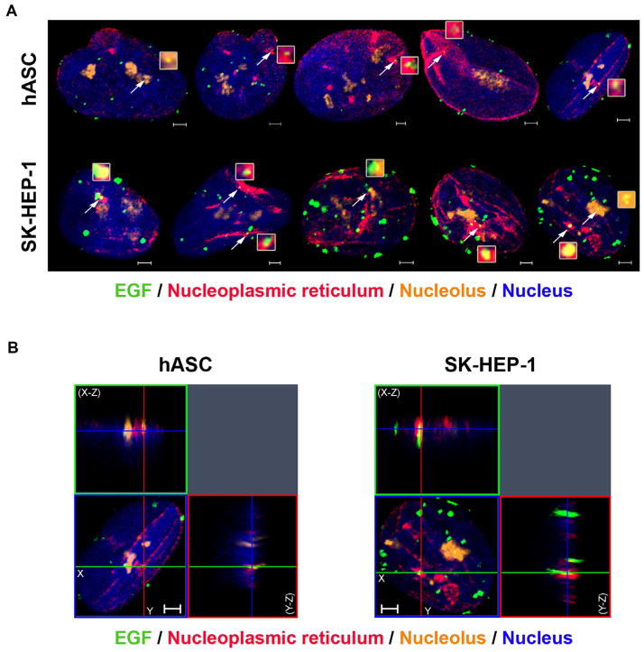
Nuclear EGF localizes in nuclear envelope invaginations and in nucleoli of both cell types. A- hASC and SK-HEP-1 cells were stimulated with EGF conjugated to Alexa Fluor® 488 for 40 min and had the nuclear envelope stained with an anti-lamin B2 antibody, the nucleolus stained with an anti-brillarin antibody and the nucleus labeled with Hoechst (in blue). The white arrows point to EGF clusters (in green) located in invaginations of the nuclear envelope (in red) and in the nucleolus (in orange). These clusters are in light yellow and in higher magnitude in the inserts next to the arrows. Scale bar: 2 μm. B- Three-dimensional reconstruction of the serial optical sections of 40 min-stimulated hASC and SK-HEP-1 cells. X–Z sections are shown at the top, and Y–Z sections are shown on the right of each image. Images are representative of 41 (hASC) and 37 (SK-HEP-1) cells. Scale bar: 2 μm. (For interpretation of the references to color in this figure legend, the reader is referred to the web version of this article.)

