
Fig. 2.
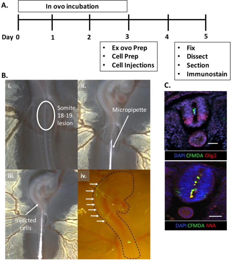
Ex ovo transplantation method. (A) Timeline of experimental protocol with (B) images depicting the (i.) midline neural tube lesion, (ii. and iii.) injection of hMN cultures, and (iv.) visualization of CFMDA dyed cells (arrows) under a stereo microscope immediately afterwards. (C) Immunostained Olig2+ spinal tissue sections (top) showing transplanted cells positive for CFMDA dye and human nuclear antigen (hNA, bottom) in the central canal. Scale bars are 50μm.