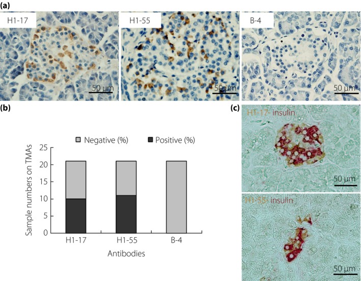
Figure 1.

Representative immunohistochemical images of two cross‐reactive antibodies in human pancreatic tissue microarrays. (a) From the staining pattern, H1‐17 and H1‐55 clones are stained with the surrounding cells of the pancreas islet. The sections were counterstained with hematoxylin (B‐4 is immunoglobulin G1 isotype antibody used as a negative staining control). (b) Positive numbers of immunohistochemical staining from 21 different pancreas tissues samples. Nearly 50% of samples were expressed in both H1‐17 and H1‐55 clones. (c) Immunohistochemical double staining of H1‐17 and H1‐55 (3,3′‐diaminobenzidine staining) with anti‐insulin (alkaline phosphatase‐red). Both antibodies are not localized in insulin‐secreting β‐cells. TMAs, tissue microarrays.
