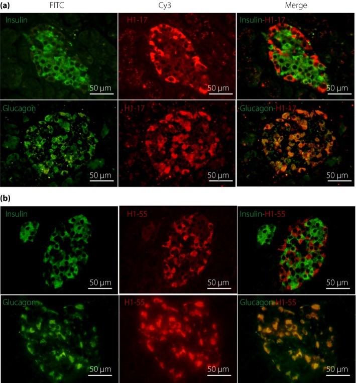
Figure 2.

Representative images of double indirect immunofluorescence of two cross‐reactive antibodies with anti‐insulin and anti‐glucagon antibodies on human pancreatic tissues. (a) H1‐17 clone; (b) H1‐55 clone. Merged images showed that H1‐17 and H1‐55 antibodies were located in the glucagon‐secreting α‐cells (yellow signal), not β‐cells. FITC, fluorescein‐isothiocyanate.
