Figure 1.
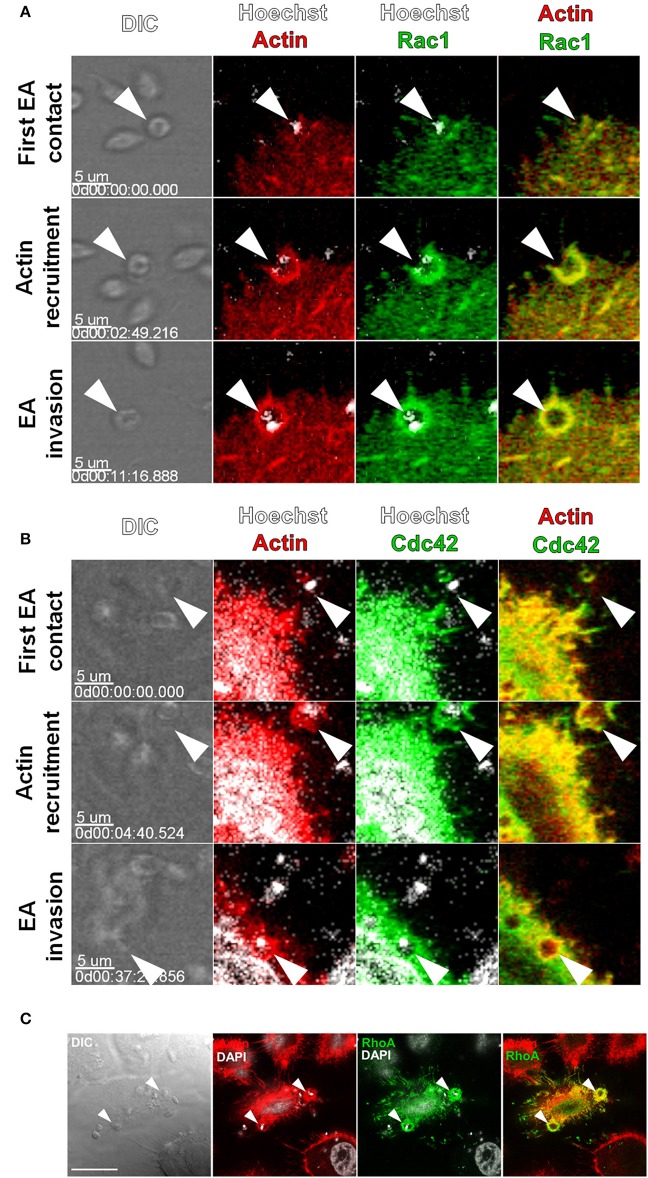
Rac1, Cdc42 and RhoA are recruited to actin-rich cup-like structures induced by EAs. HeLa cells were transfected with Cdc42, Rac1 (GFP tagged) or RhoA (c-myc tagged) and interactions with EAs were evaluated by live confocal microscopy. (A) Frames from time-lapse movies showing the moments of initial contact, beginning of actin recruitment and EA (arrowheads) internalization with recruitment and colocalization of Rac1-GFP (green) and LifeAct-RFP (red). (B) Frames from time-lapse movies showing Cdc42-GFP (green) recruited along with LifeAct-RFP (red) to EA (arrowheads) invasion sites during internalization. (C) Recruitment and colocalization of RhoA-c-myc (green) with actin (red) at EA invasion sites (arrowheads) was observed in fixed cells stained with phalloidin-TRITC. Nuclei and kinetoplasts were stained with Hoechst (A,B) or DAPI (C). Bar = 20 μm.

