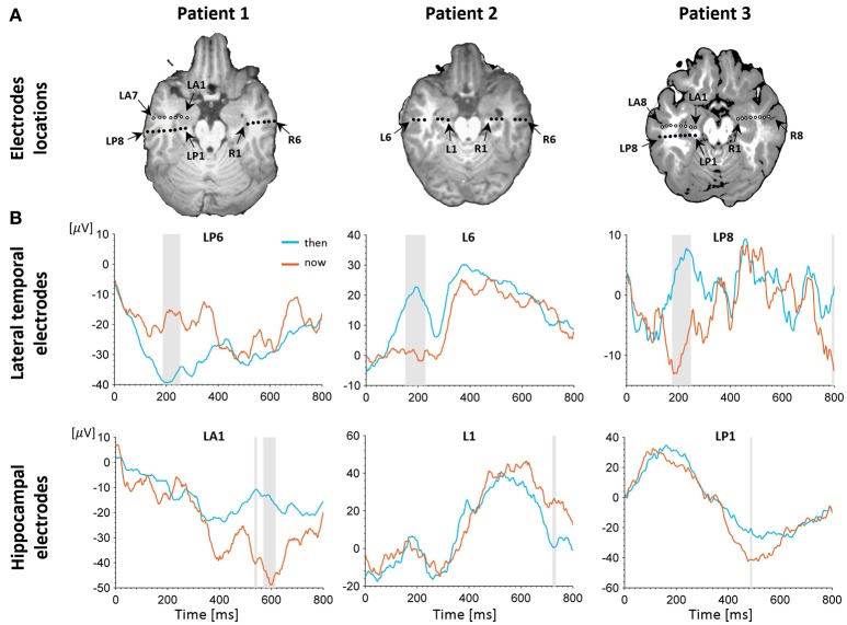
Figure 2.
Electrophysiological results. (A) Depth electrodes locations in the hippocampus and lateral temporal cortex (LTC), shown for each patient on a co-registration of post-operative CT scan and pre-operative MRI (white circles depict electrodes projected on this slice for visualization purposes; for more precise localization of these electrodes see Figure S2. Exact neuroanatomical position of each electrode as verified by two certified neuro-radiologists is available in Table S1. (B) Intracranial evoked potentials (iEPs) recorded at representative electrodes in the left LTC (top) and left hippocampus (bottom) during MTT. LTC electrodes show high early task modulation, whereas electrodes in the hippocampus show high late task modulation. Shaded areas show time points of significant differences between conditions in a two-tailed independent samples t-test (p < 0.05, uncorrected).

