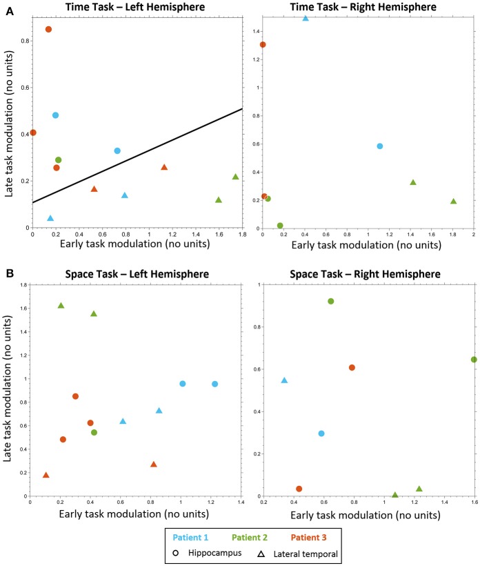
Figure 3.
Electrodes classification. Electrodes classification using linear SVM, based on early task modulation value (X-axis) and late task modulation value (Y-axis). (A) MTT task: left hippocampal electrodes (circles) are clearly separable from left lateral temporal cortex (LTC) electrodes (triangles) on the plane of early and late task modulations (see Figure S3 and Table S2). A separating line is shown, as obtained from SVM classification of all electrodes (left, p < 0.005). No such separation was found for electrodes in the right hemisphere (right, see Figure S4). (B) Spatial task: no separation between lateral temporal and hippocampal electrodes was found neither in the left hemisphere (left, see Figure S6) nor in the right (right, see Figure S7).

