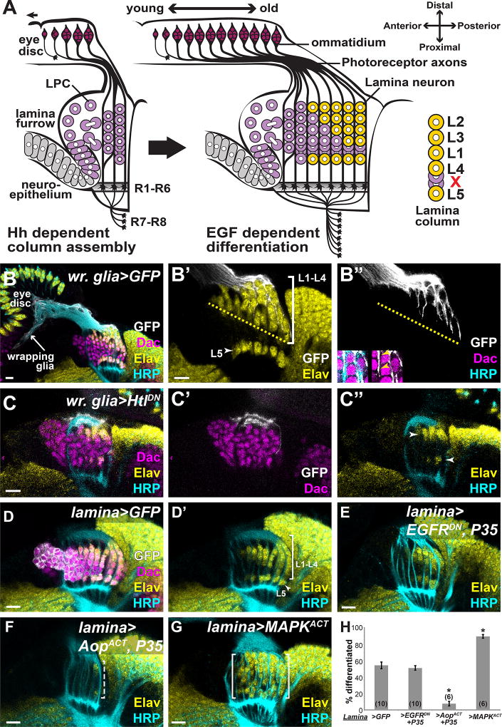
Fig. 1. Photoreceptors do not communicate directly with lamina precursors through EGF.
(A) Schematic of lamina development in the optic lobes, which is coupled to photoreceptor development in the eye disc. Hh from photoreceptors drives lamina precursor (purple) birth and assembly into columns. Photoreceptor-EGF is required for precursor differentiation into neurons (yellow). Columns consist of 6–7 precursors, which differentiate in an invariant spatiotemporal pattern (yellow). (B) A horizontal view of an early pupal (P10–15hrs APF) eye disc and optic lobe showing photoreceptor axons marked by HRP (cyan). In the optic lobe, lamina precursors express Dac (magenta) and differentiated photoreceptors and neurons express Elav (yellow). Lamina cell bodies (magenta) are organized into columns that associate with photoreceptor axons. Wrapping glia, marked by membrane-targeted GFP (white) driven by a wrapping glia-specific Gal4, extended processes through the optic stalk and into the lamina, where they encapsulate lamina cells and photoreceptors progressively (inset in B”; arrowheads mark location of photoreceptors between glial processes and lamina cells). (C) Expressing HtlDN in wrapping glia disrupted glial process infiltration into the lamina. Only cells immediately below glial processes differentiated (arrowhead in D”). (D) Lamina-specific Gal4 driving GFP showed normal lamina neuron differentiation. (E) Lamina-specific EGFRDN and P35 co-expression did not affect neuronal differentiation. (F) Lamina-specific AopACT and P35 co-expression led to loss of differentiated neurons (dashed bracket). (G) Lamina-specific MAPKACT expression led to premature Elav expression in columns. (H) Quantification of (E–F) as a percentage of differentiated cells in the 6 youngest lamina columns. Asterisks indicate significance with Mann-Whitney U-test p<0.01; #optic lobes examined indicated in brackets. (Scale bar = 10µm).

