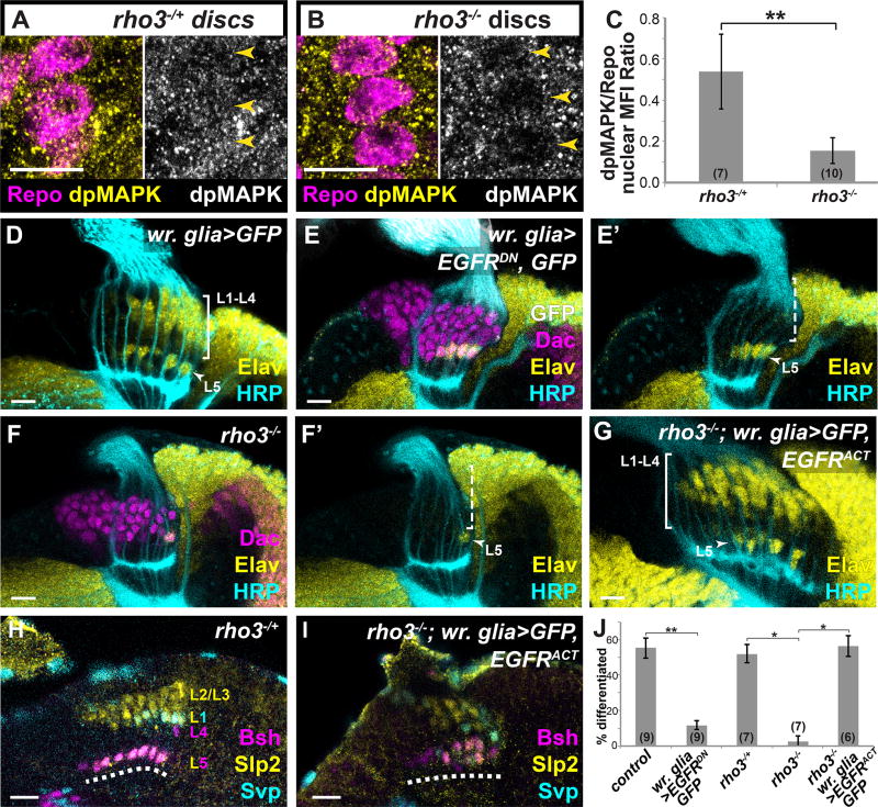
Fig. 2. L1–L4 differentiation requires photoreceptor-induced EGFR signaling in wrapping glia.
Eye discs with wrapping glia marked by the pan-glial nuclear marker Repo (Magenta) and dpMAPK (yellow) in (A) rho3−/+ and (B) rho3−/− animals, quantified in (C) p<0.001; Mann-Whitney U-test; #discs indicated in brackets. (D–G) Optic lobes stained for Elav (yellow), Dac (magenta), HRP (cyan) and GFP (white) (D) A control wr. glia>GFP lamina. (E) When wrapping glia express EGFRDN, only presumptive L5s differentiated (arrow head). (F) In a rho3−/− animal, there was only a late differentiating presumptive L5 (See also Fig. S1C–G). (G) When wrapping glia express EGFRACT and GFP in a rho3−/− background, the L1–L4 front of differentiation is restored (bracket). (H,I) Developmentally expressed subtype-specific markers used in combination to identify neuronal subtypes (16): Sloppy paired 2 (Slp2) alone marks L2 and L3; Slp2 and Seven up (Svp) together mark L1, Brain-specific homeobox (Bsh) alone marks L4, and Slp2 and Bsh together mark L5 (dashed line indicates lamina plexus). (H) In a control rho3−/+ brain and (I) when wrapping glia drive EGFRACT (and GFP; not shown) in a rho3−/− background, all cell types were recovered. (J) Quantification of (D–G) as a percentage of differentiated cells in the 6 youngest lamina columns. Asterisks indicate significance with Mann-Whitney U-test p<0.01; #optic lobes examined indicated in brackets. (Scale bar = 10µm).

