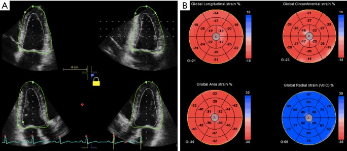
Figure 2.
3D strain parameters of the left ventricle in a healthy subject. From a single end-systolic ROI (A), longitudinal, circumferential and area strain (negative—red color) and radial strain (positive—blue color) are simultaneously obtained (B); in the left bottom corner of each bull’s eye map, the global value is displayed for each strain parameter (G, global).

