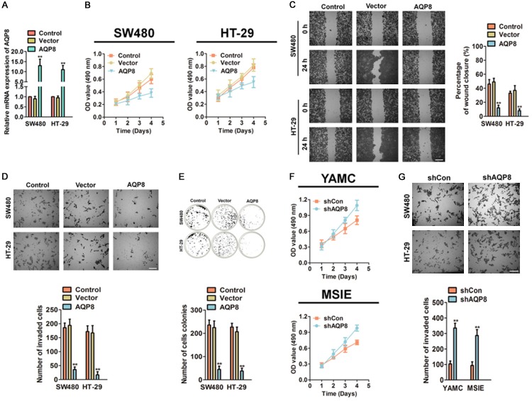
Figure 2.
Over-expression of AQP8 inhibits cells growth, invasion and colony formation. A. SW480 and HT-29 cells were transfected with pcDNA4-myc/his-AQP8 or pcDNA4-myc/his vector and the mRNA of AQP8 were analyzed by qPCR. B. Cell proliferation of control CRC cells and AQP8 over-expressing cells was determined by MTT analysis. C. The mobility of AQP8 over-expressing cells was assessed by wound healing analysis. Scale bar: 200 μm. D. Boyden invasion assay was conducted using control cells and CRC cells transfected with AQP8. Scale bar: 200 μm. E. Colony formation assay was conducted to evaluate the anchorage-independent growth of indicated cells. **P < 0.01 compared to control cells. F. AQP8 knocked-down accelerated cell proliferation in YAMC and MSIE cells as shown in MTT analysis. G. Cell invasion was determined by Boyden invasion assay using AQP8 knocked-down YAMC and MSIE cells. Scale bar: 200 μm. **P < 0.01 compared to cells transfected with shCon.

