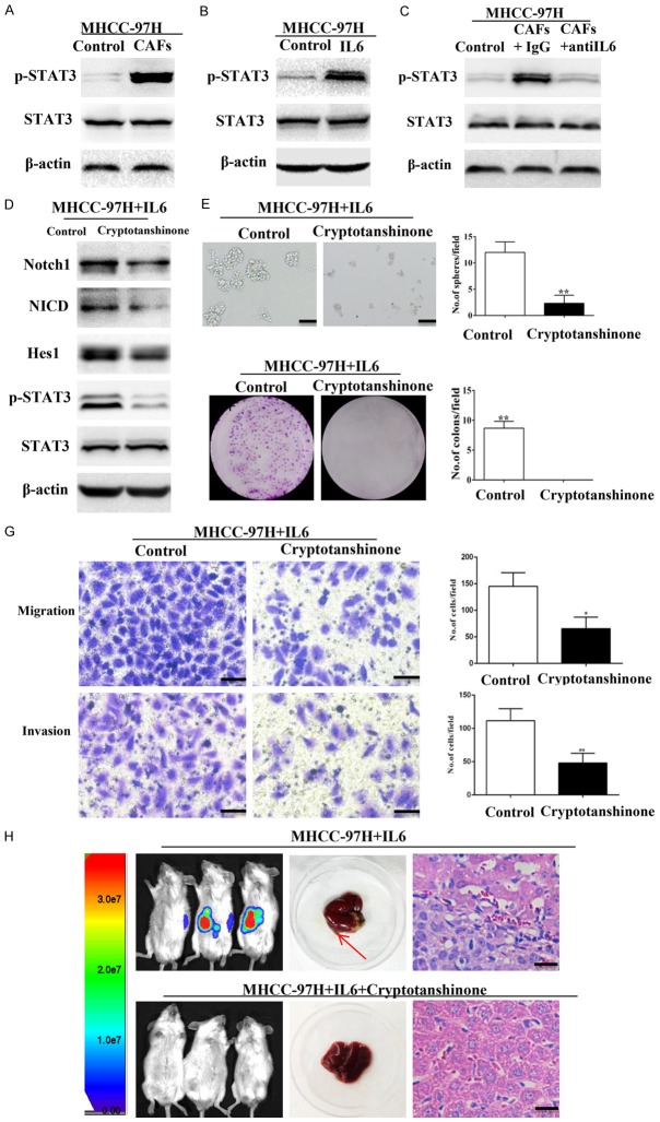
Figure 7.
IL-6 induces the activation of Notch signaling via STAT3 Tyr705 phosphorylation. (A, B) Phospho-STAT3 (Tyr705) was elevated in MHCC-97H cells treated with CAFs (A) or IL-6 (B). (C) IL-6-neutralizing antibody abolished CAFs-induced overexpression of Phospho-STAT3 (Tyr705) in MHCC-97H cells. (D) Cryptotanshinone suppressed the expression of phospho-STAT3 and Notch signaling components (Notch1, NICD, and Hes1) in MHCC-97H cells treated with IL-6. (E, G) IL-6-induced sphere-forming (E), colony-forming (F), and migration and invasion (G) abilities of MHCC-97H cells decreased when STAT3 Tyr705 phosphorylation was inhibited by cryptotanshinone. Scale bar, 100 μm. (H) MHCC-97H cells treated with IL-6 in the presence or absence of cryptotanshinone were injected into the livers of NOD/SCID mice. Inhibition of STAT3 Tyr705 phosphorylation abolished the effect of IL-6. Paraffin-embedded tissues of xenotransplanted tumours were processed for H&E staining. Scale bar, 50 μm. Red arrow indicates the site of tumor formation of MHCC-97H cells in the livers of NOD/SCID mice. Data are shown as means ± SD from at least three independent experiments. (*P < 0.05, **P < 0.01).

