Figure 4.
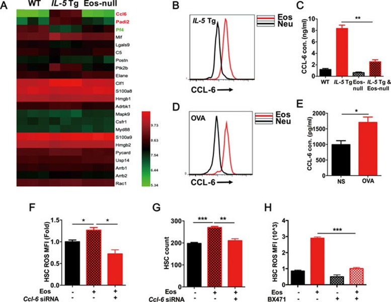
Eos-derived CCL-6 is responsible for disrupted HSC homeostasis. (A) Heatmap of screened cytokines and chemokines in bone marrow supernatants of WT, IL-5 Tg, Eos-null mice analyzed by mass spectroscopy. (B) CCL-6 MFI analysis by FACS in Eos and Neu from IL-5 Tg mice. (C) CCL-6 levels determined by ELISA in the serum of WT, IL-5 Tg, Eos-null and IL-5 Tg and Eos-null mice. Cons, concentrations. (D) CCL-6 MFI analysis by FACS in Eos and Neu from OVA-challenged and control mice. (E) CCL-6 levels determined by ELISA in the serum of OVA-challenged and control mice. (F) ROS MFI analysis in LT-HSCs co-cultured with Eos transfected with negative control siRNA (NC) or Ccl-6 siRNA for 2 h. (G) HSC migration assay after co-culture for 12 h with Eos transfected with NC or Ccl-6 siRNA. (H) ROS MFI analysis in HSCs co-cultured for 2 h with Eos following treatment with BX471. Data are shown as the means ± SEM with six samples per group. *P < 0.05, **P < 0.01, ***P < 0.001 versus respective controls.

