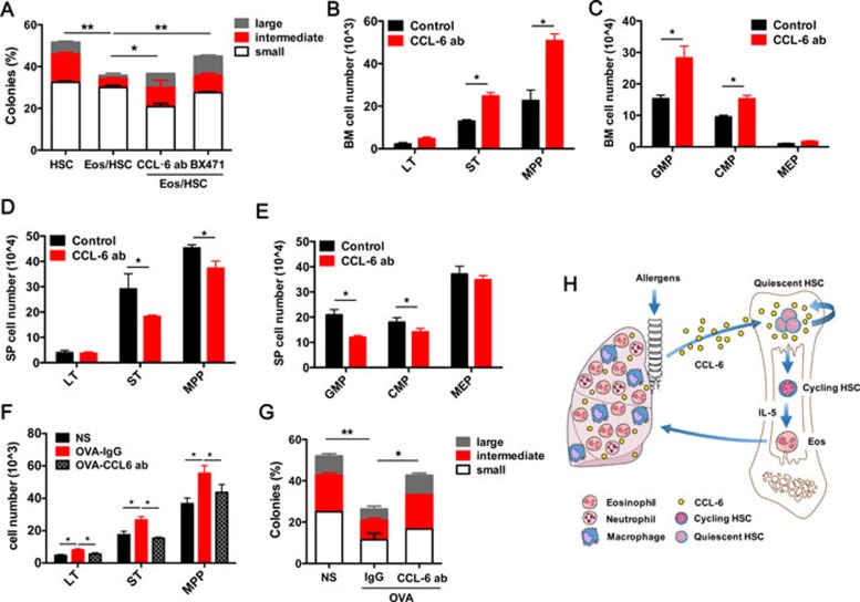
Figure 5.
Neutralizing of CCL-6 in IL-5 Tg and OVA-treated mice rescue HSC impairment. (A) Percentage of large, intermediate and small colonies in the single-colony forming assay of sorted LT-HSCs co-cultured with Eos and intervened with the CCL-6 antibody and BX471 (n = 3). (B, C) Absolute numbers of stem cells (B) and progenitor cells (C) in the BM of IL-5 Tg and control mice treated with CCL-6-neutralizing antibody. (D, E) Absolute numbers of stem cells (D) and progenitor cells (E) in the SP of IL-5 Tg and control mice treated with CCL-6 antibody. (F) Absolute numbers of stem cells in the BM of OVA-challenged mice treated with CCL-6-neutralizing antibody. (G) Percentage of large, intermediate and small colonies in a single-colony forming assay performed on LT-HSCs sorted from OVA-challenged mice treated with CCL-6-neutralizing antibody. (H) Schematic representation of the roles of Eos and Eos-secreted CCL-6 in regulating HSC function in allergen-induced airway inflammation. Data are shown as the means ± SEM with six samples per group. *P < 0.05, **P < 0.01 versus respective controls.

