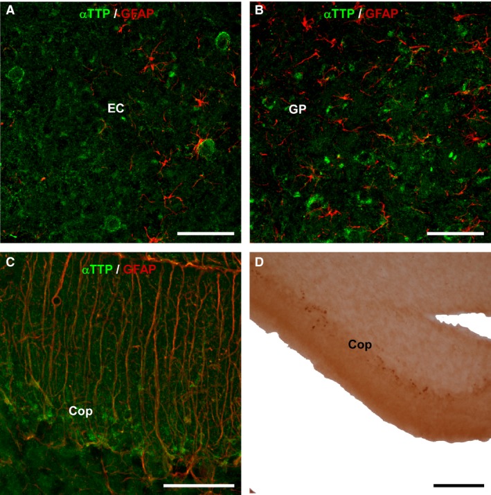
Figure 2.

Double immunohistochemistry for GFAP/αTTP. The images were obtained using laser confocal microscopy and show double immunohistochemistry for GFAP (red) and αTTP (green) in 24‐month‐old KO mice. (A) Sagittal section showing the entorhinal cortex (EC). (B) Globus pallidus. (C) The Purkinje cell layer of the cerebellum contained some double‐labeled cells that might represent Bergmann's glia. Otherwise, no double labeling was detected. (D) Coronal section of the cerebellum showing single labeling for αTTP (18 months old, heterozygote). Thick points can be observed in the layer of Purkinje cells. Scale bars: 50 μm (A, B, C), 100 μm (D). CoP, cerebellum, layer of Purkinje cells; EC, entorhinal cortex; GP, globus pallidus.
