
Figure 4.
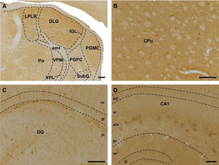
Immunolabeling for SVCT2. (A) Coronal section of thalamic nuclei (wt, 18 months old). (B) Immunoreactivity for SVCT2 in the caudate and putamen (wt, 4 months old). (C) Sagittal section showing the different layers of the dentate gyrus, in which cell bodies are located mainly in the polymorphic layer (wt, 4 months old). (D) Sagittal section of CA1, in which thick SVCT2‐positive profiles arranged in clusters are located in the stratum lacunosum moleculare (24 months old, KO). Scale bar: 100 μm. CA1, Cornus Ammonis, Ammon's horn (field of the hippocampus); CPu, caudate putamen; DG, dentate gyrus; DLG, dorsal lateral geniculate nucleus; eml, external medullary lamina; gl, granular layer; hf, hippocampal fissure; IGL, intergeniculate leaflet; LPLR, lateral posterior thalamic nucleus; ml, molecular layer; PGMC, pregeniculate nucleus, magnocellular part; PGPC, pregeniculate nucleus, parvocellular part; pl, polymorphic layer; Po, posterior thalamic nuclear group; pyl, pyramidal layer; slm, stratum lacunosum‐moleculare; sr, stratum radiatum; SubG, superficial gray layer of the superior colliculus; VPL, ventral posterolateral thalamic nucleus; VPM, ventral posteriomedial thalamic nucleus.