Figure 7.

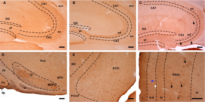
Double‐immunostaining for SVCT2 and αTTP. (A) SVCT2‐immunoreactive profiles in the hippocampal formation (heterozygote, 4 months old). (B) Mossy fibers displayed TTPα‐immunoreactivity (wt, 24 months old). (C) Coronal section of the hippocampus, in which SVTC2 (brown precipitate) and αTTP (blue color) were observed (wt, 24 months old). Arrowheads: SVCT2‐positive neurons. (D) Sagittal section of the brainstem displaying the trapezoid body (tz), which was positive for αTTP (blue color), and the nucleus of the trapezoid body (TZ), which contained SVCT2‐immunoreactive (brown precipitate) neurons (heterozygote, 4 months old). (E) Sagittal section of the inferior colliculus showing neurons exhibiting double immunostaining (wt, 24 months old). (F) Coronal section of the granular part of the retrosplenial cortex, in which pericellular immunostaining for SVCT2 (white arrowhead) and αTTP (blue arrowhead) as well as double‐labeled cells (black arrowheads) were observed (wt, 24 months). Scale bar: 100 μm. I‐VI, cortical layers; BIC, nucleus of the brachium of the inferior colliculus; CA1 and CA3, Cornus Ammonis, Ammon's horn (fields of the hippocampus); DG, dentate gyrus; ECIC, external cortex of the inferior colliculus; mf, mossy fibers; MVPO, medioventral periolivary nucleus; pl, Polymorphic layer; Pn, pontine nuclei; PnC, pontine reticular nucleus; PrCnf, precuneiform area; py, pyramidal tract; RSGc, retrosplenial granular cortex, c region; scc, splenium of the corpus callosum; so, stratum oriens; SPO, superior paraolivary nucleus; tfp, transverse fibers of the pons; TZ, nucleus of the trapezoid body; tz, trapezoid body.
