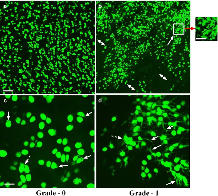
Figure 2.

Low and high power axial confocal laser scanning microscope reconstructed images of chondrocytes within grade‐0 and grade‐1 human femoral head articular cartilage. Representative examples of confocal laser scanning microscope images at low magnification (×10) 5‐chloromethylfluorescein diacetate‐ and propidium iodide‐labelled chondrocytes (live and dead cells, respectively) in human (a) grade‐0 and (b) grade‐1 cartilage explants. At low magnification, chondrocyte morphology and distribution in grade‐0 cartilage appeared relatively normal; however, in mildly degenerate (grade‐1) samples, evidence of chondrocyte clustering (solid arrow) and areas of hypocellularity were present (double‐headed arrows). Scale bars: 100 μm (a,b) and 50 μm (inset). At high magnification (panel c) (×40), the majority of chondrocytes in grade‐0 cartilage demonstrated normal morphology, but some cells with short, often curled, cytoplasmic processes were present (solid arrows) with infrequent clustering of chondrocytes (≥ 3 cells; broken arrow). In grade‐1 cartilage (d), many cells possessed cytoplasmic processes of varying length (solid arrows) and number, and there was extensive clustering (inset in b, and broken arrow in d). Note that there were no propidium iodide‐labelled chondrocytes detected in these images even though this fluorescent indicator was present. Scale bars: 25 μm (c,d).
