Figure 1.
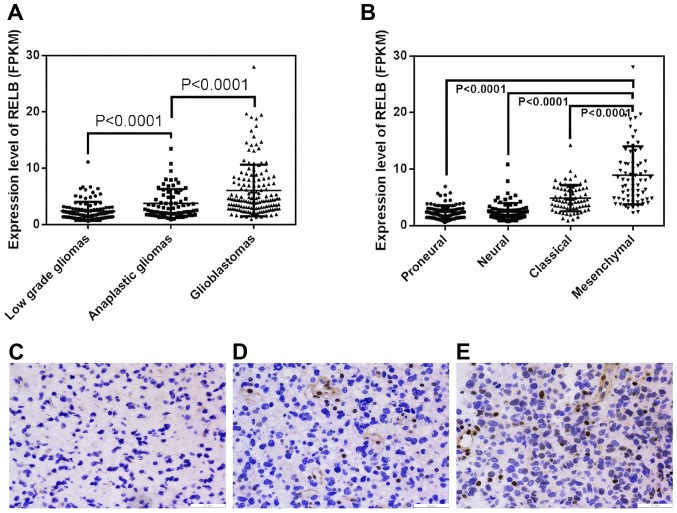
The expression difference of relB in different grade and pathologcal type gliomas. (A, B) A single spot was the expression value of relB of an individual patient. Lines in the middle were the mean expression value (A and B). Immunocytochemical staining of relB in different grade tumor tissues (mgnification, ×200) (C) low grade gliomas, (D) anaplastic gliomas and (E) glioblastomas.

