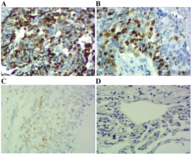
Figure 3.
Immunohistochemical staining of squamous cell carcinoma tissue sections demonstrating p-STAT3 expression (magnification, ×200). (A) Squamous cell carcinoma specimen demonstrating high expression of p-STAT3-positive tumor cells (score 3). (B) Squamous cell carcinoma specimen demonstrating moderate expression of p-STAT3-positive tumor cells (score 2). (C) Squamous cell carcinoma specimen demonstrating low expression of p-STAT3-positive tumor cells (score 1). (D) Photomicrographs of a corresponding normal lung tissue specimen with no p-STAT3-positive tumor cells (score 0). p-STAT3; phosphorylated signal transducer and activator of transcription 3.

