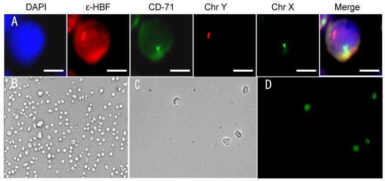
Figure 5.
(A) Identification of fNRBC in maternal peripheral blood using immunofluorescent staining and FISH technique. Representative pictures of fNRBCs from gravida who conceived male infants are shown. The scale bars are 5 μm. (B-C) Representative images of nucleated red blood cells on the microchip before/after electric stimulation by 0.8 V. (D) FDA and PI staining of cells on the microchip after 0.8 V electric stimulation.

