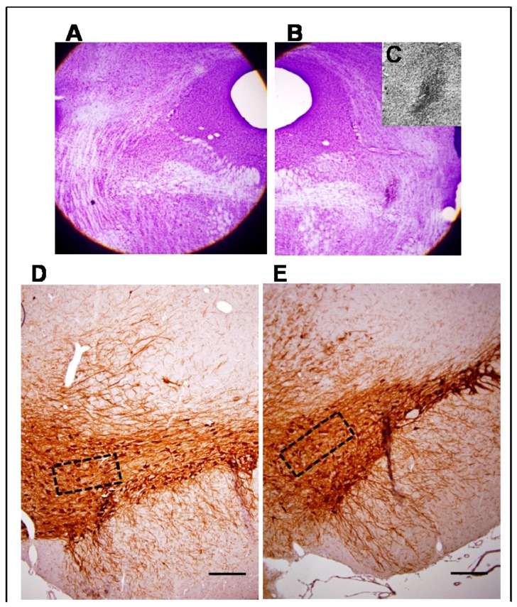
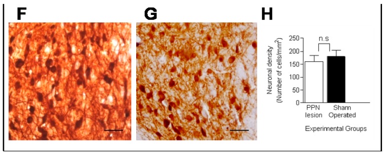
Figure 1.
Morphological study. (A,B) Composition made from photomicrographs of brain coronal sections of a seven-day lesioned rat. (A) PPN area from the left side, contralateral to the lesion; (B) Cresyl Violet study revealed the zone of injury in the area occupied by the right PPN adjacent to the superior cerebellar peduncle (4×); (C) Magnification of the lesion area (10×); (D–G) Representative photomicrographs of the immunohistochemical study for tyrosine hydroxylase in nigral coronal sections of: Sham-operated (D) and NMDA-lesioned rats (E) (4×). The area within the rectangle of each image is magnified (40×) in (F) and (G) respectively. Note that dopaminergic cell bodies are well conserved in the SNpc on both sides; (H) Comparison of neuronal density showed non-significant differences between the right SNpc from NMDA-lesioned rats and Sham-operated rats groups (n = 5 for each experimental groups). (Scale bar for D and E = 100 µm; scale bar for F and G = 50 µm).


